veterinary reports
Information
The complete examination documents and reports can be sent by email. Examinations were taken by our regular veterinarian, an orthopaedic specialist, two neurologists from the Zurich University Veterinary Hospital and a neurologist from the Marigin Veterinary Clinic.
Zurich University Veterinary Hospital
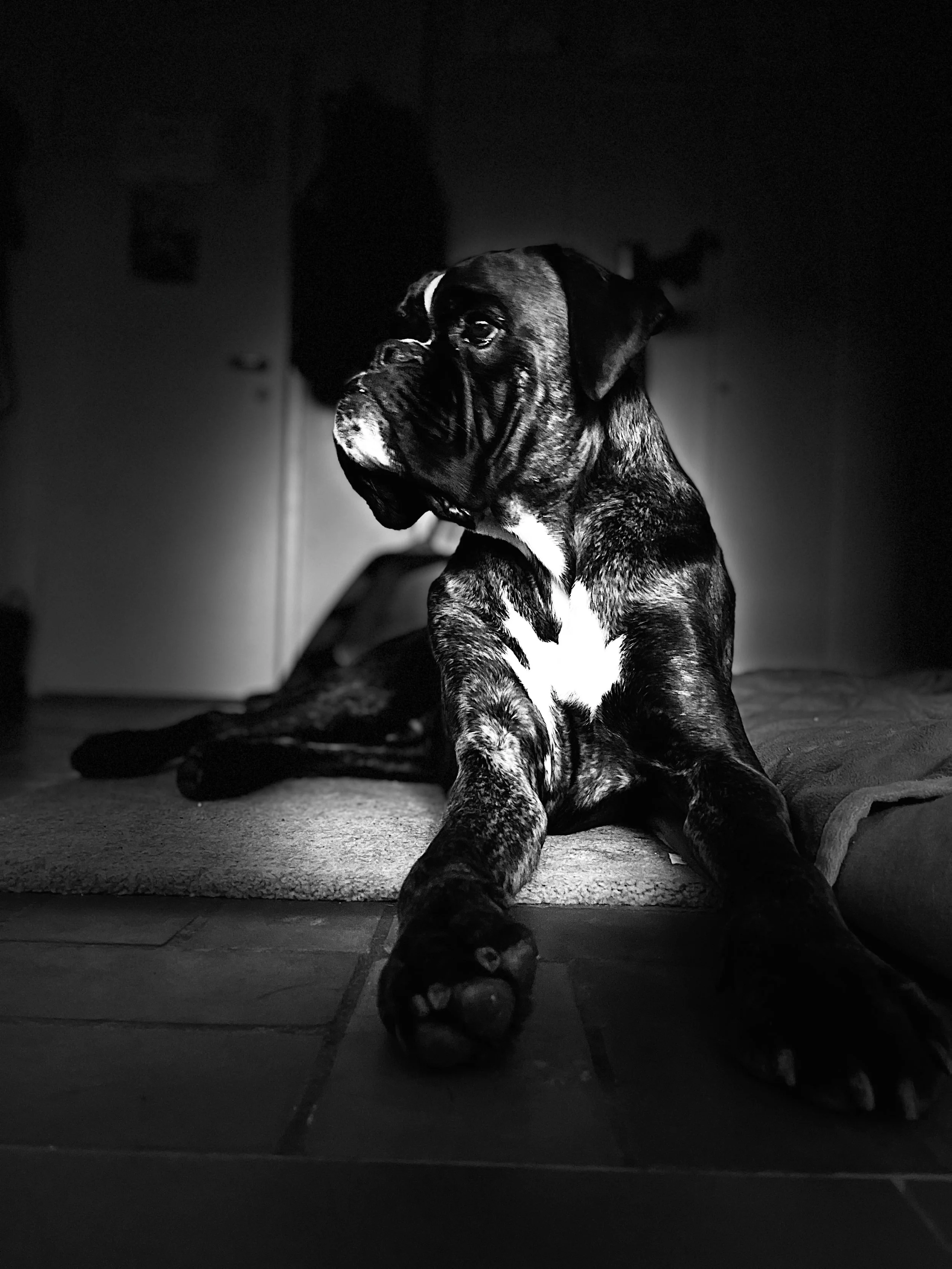
Clinical history of Merlot - Boxer - 4 years old - male - neutered
Dog was presented in September for left side front limb lameness. At this stage MRI of the neck and the brachial plexus was performed and no changes were found. EMG at this stage EMG showed bilateral spontaneous activity in both distal front limbs. The dog has proceeded to a non ambulatory tetrapareses, more severe in the front limbs.
A repeated MRI scan was still unremarkable, EMG now also shows changes in the hind limbs. Ulnar and radial nerve could not be stimmulated. The nerve conuction velosity in the distal tibial nerve was mildly reduced (43 m/s, without any polyphasia, Proximal NCV 78 m/s). Polyneuropathy (more distal).
Comment Neurology
Merlot was presented for reevaluation and further clarification of a progressive paralysis of both front legs: Therapy with high-dose steroids had shown no improvement.
The MRI showed no evidence of involvement of the central nervous system. The proximal part of the nerves (brachial plexus) also showed no morphological changes.
Electrodiagnostics revealed mild changes in the hind legs and severe changes in the forelegs. In the area of the front legs, both the radial nerve and the ulnar nerve on the right were so severely affected that nerve conduction (nerve impulse) could no longer be triggered. The findings in the musculature are consistent with a high degree of denervation.
The biopsy findings show a severely damaged nerve with no signs of inflammation.
The cause of this massive neuropathy remains unclear. Unfortunately, it is to be feared that the damage will continue to progress. All that remains is supportive therapy using physiotherapy and the hope that the process could be self-limiting.
Institute of Veterinary Pathology
Nerve sample 12 mm long and 3 mm thick with no evident macroscopic changes.
HISTOLOGICAL FINDINGS
Sections were stained with the haematoxylin eosin (HE) stain unless stated otherwise.
Fibroadipose tissue (Nerve): The sample consists entirely of fibroadipose tissue with two highly cellular central bundles surrounded by a fibrous band (anatomically compatible with nerve samples surrounded by epineurium). The axons and myelin sheets are not recognisable and the bundles are completely composed of eosinophilic spindle cells with elongated nuclei.
Toluidine blue (semithin section): Axons and myelin sheets are not recognisable. Scattered in the endoneurium are some macrophages with large cytoplasm sometimes containing intensely
H23-3022 / page 1/2
blue and concentrically organised lamellar material (morphologically compatible with myelino- phages).
IMMUNOHISTOCHEMICAL FINDINGS
Immunohistochemistry for Periaxin (Schwann cells marker) and Neurofilament (axon marker) has been performed on 22.12.2023. Scattered throughout the sample are very rare periaxin-positive cells (Schwann cells) and very rare neurofilament-positive elements (axons).
DIAGNOSIS and COMMENT
Diagnosis:
- Fibroadipose tissue (Nerve): Severe, diffuse, chronic axon and myelin sheets loss
Comment:
The histological appearance of the sample is highly unusual and difficult to interpret. Based on the anatomical localisation reported for the submitted specimen and the presence of bundles surrounded by a fascia that could be compatible with epineurium, the samples were interpreted as nerves. However, it is not possible to clearly identify axons or myelin sheets on haematoxylin-eosin stains. The semi-thin section stained with toluidine blue could indicate the presence of myelinophages, suggesting a nerve damage (primary or secondary). Immunohistochemistry showed only rare elements compatible with Schwann cells and axons. The main hypothesis is that the submitted nerve (right n. ulnaris) has lost its axons and associated Schwann cells, leaving only the connective tissue scaffold. The second hypothesis is that a structure such as a ligament or tendon was accidentally sampled. However, the high cellularity is unusual for such structures as well as the presence of rare periaxin-positive cells (Schwann cells) or neurofilament-positive elements (axons).
MRI
MRI examination of the head, the cervical and cranial thoracic spine and brachial plexus, skeletaly mature dog. MRI examination of the 25.9.23 as a comparison available.
Head
Surrounding musculature and calvarium unremarkable.
Cerebral hemispheres with unremarkable sucli and gyri. Definition of white and grey matter maintained.
Lateral ventricles symmertrically and bilaterally mildly widened.
Caudal part of the septum pellucidum not delineated.
Liquor with normal T2 signal intensity.
Brainstem und cerebellum unremarkable.
Cervical spine and brachial plexus region
Mildly enlarged crevical superficila lymph nodes, L>R, unchanged.
Shoulder joints only partially included in the study, unchanged.
Bony structures of the cervical and thoracic spine unremarkable.
INtervertebral discs: nuclei pulposi with normal volume and T2 signal intensity, no compressive lesions.
The spinal cord and the central canal are unremarkable.
Brachial plexus bilaterally unremarkable.
Mild bilateral muscle atrophy at the level of the shoulders.